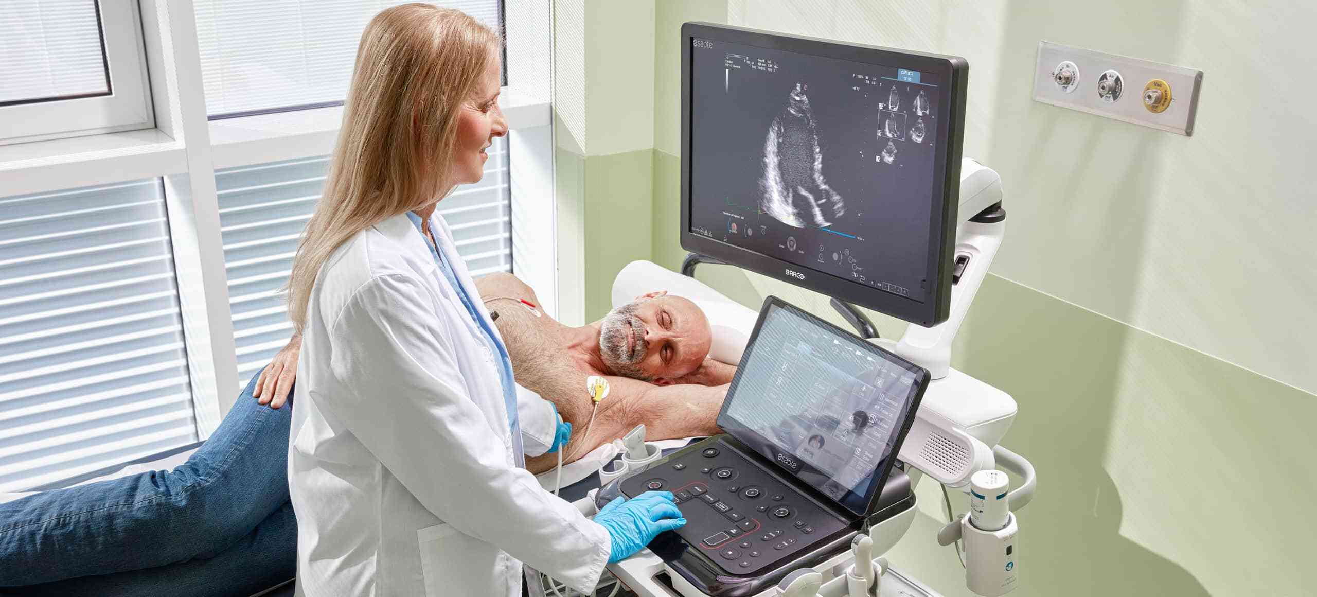
Ultrasound clinical solutions for Cardiovascular
Echocardiography remains the cornerstone of cardiovascular diagnostics—non-invasive, real-time, and essential for evaluating cardiac structure, function, and hemodynamics. From the initial diagnosis of heart failure to the ongoing monitoring of cardiomyopathies, valvular diseases, and ischemic conditions, the quality and efficiency of echocardiographic assessment directly impact patient outcomes.
As clinical workloads increase and patient cases become more complex, cardiologists need solutions that are not only accurate and rapid but also standardized, reproducible, and easy to integrate into daily practice.
Esaote’s advanced cardiac ultrasound suite meets this demand with a comprehensive portfolio of intelligent tools that transform routine echocardiographic exams into efficient, high-value diagnostic workflows.
INTERVIEWS
Testimonials

![]() VIDEO
VIDEO
The role of Artificial Intelligence in the echocardiography workflow
Prof. Luigi P. Badano
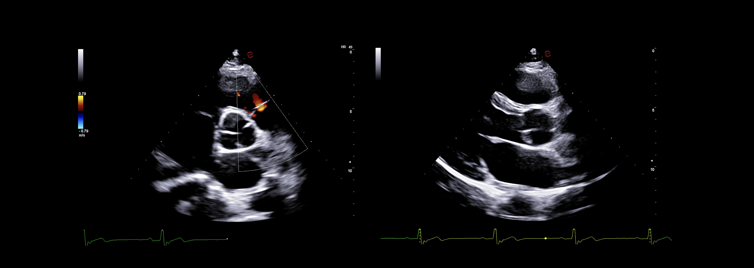
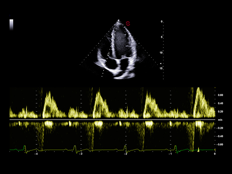
Outstanding image quality
Esaote’s XCrystal probe PX1-5 delivers exceptional clarity in B-mode imaging, providing deep penetration and excellent resolution even in technically challenging patients. Its advanced architecture ensures high Doppler sensitivity and spatial detail, enabling an accurate assessment of wall motion, valve morphology, and flow dynamics. From subtle endocardial borders to low-velocity diastolic flows, the PX1-5 probe ensures you never miss the details that matter.
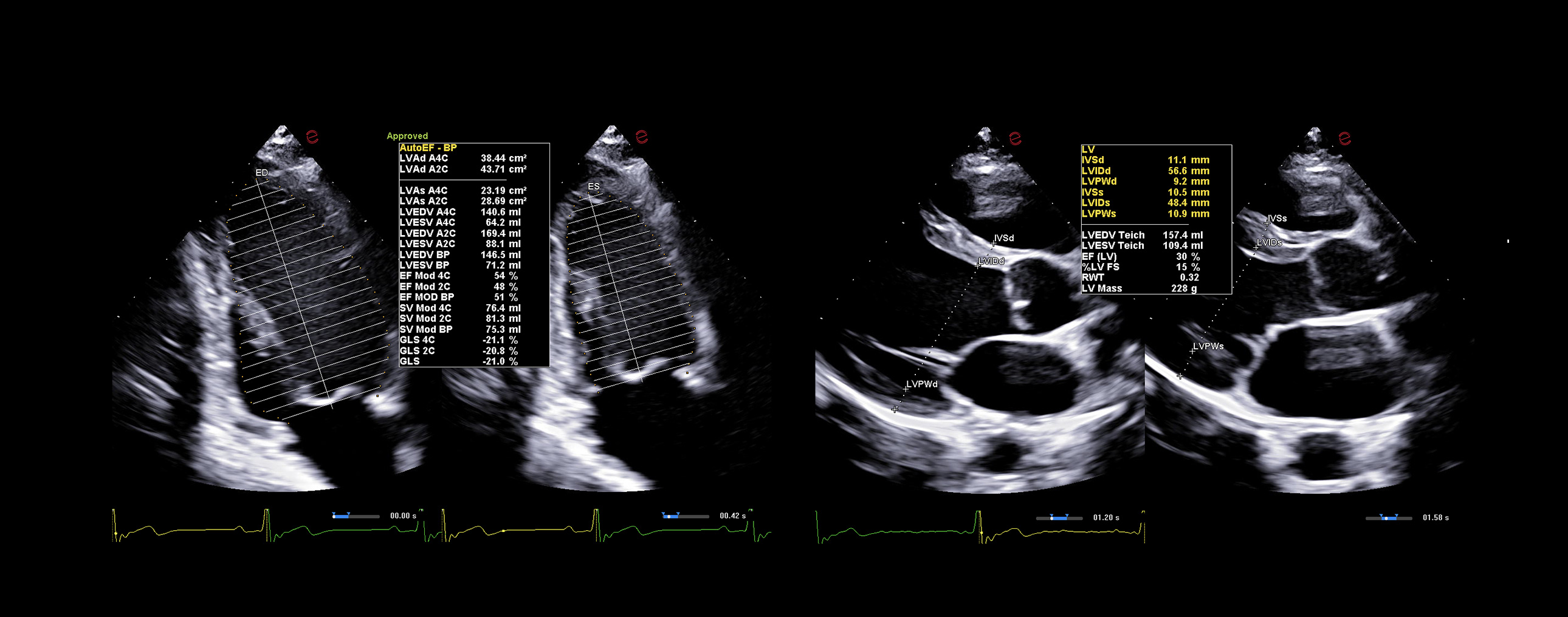
AI-based cardiac measurements for speed and consistency
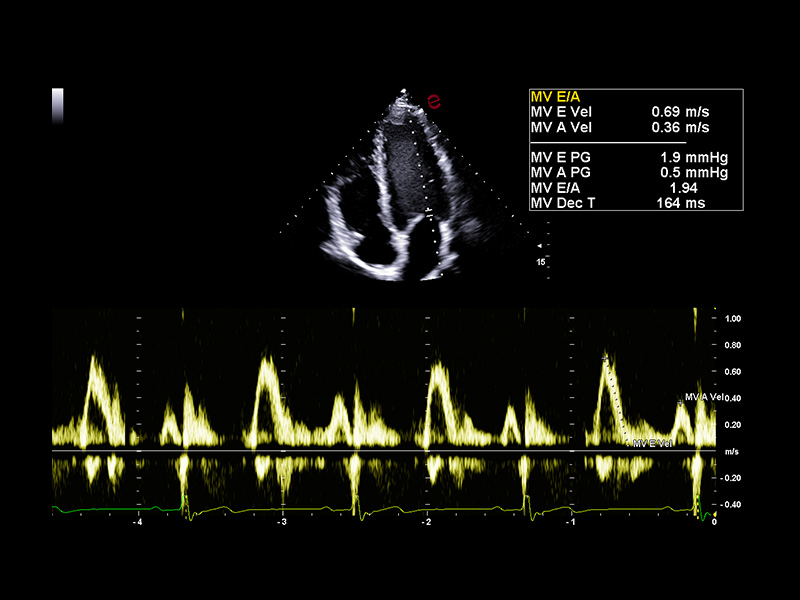
Esaote’s AutoEF and AutoCM tools are designed to bring a step ahead in the device use experience. With advanced AI algorithms, AutoEF automatically recognizes apical views and traces endocardial borders offering a proposal of left ventricular volumes and ejection fraction in just seconds. AutoCM complements this by automatically suggesting ventricular wall thickness, internal diameters, and derived values such as LV mass and fractional shortening directly from parasternal views. Moreover, AutoCM assists cardiologists by automatically recommending a calculation of the E/A wave ratio of the mitral valve, providing crucial information on diastolic function.
Together, these tools help clinicians to obtain accurate, guideline-aligned measurements in a fast way, maintaining diagnostic evidence at the top. AutoEF and AutoCM automate key functional and structural measurements—such as ejection fraction, LV volumes, wall thickness, and LV mass— making a smooth workflow, especially in high-throughput clinics.
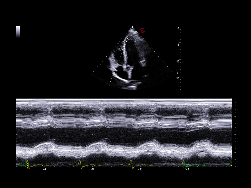
Complete and customizable cardiac measurement Suite
Esaote’s cardiac ultrasound systems provide a full range of standard measurements for structural, functional, and hemodynamic evaluations, aligning with ASE/EACVI guidelines. From M-mode and 2D linear measurements to Doppler parameters, chamber quantification, and strain analysis, all tools are fully integrated for routine clinical use. Additionally, the platform supports customizable protocols and report templates, allowing clinicians to tailor workflows to specific patient populations, exam types, or institutional standards, thereby ensuring both diagnostic consistency and operational efficiency.
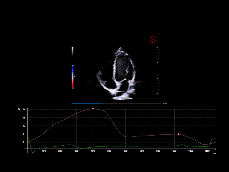
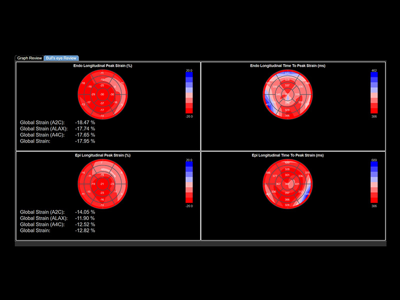
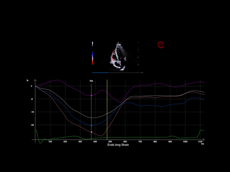
Advanced myocardial strain analysis with XStrain™
Esaote’s XStrain™ technology offers cutting-edge myocardial deformation analysis across multiple cardiac chambers and views—including the left ventricle (LV), right ventricle (RV), left atrium (LA), and short-axis (SAX) planes. Utilizing speckle-tracking algorithms, XStrain™ provides a detailed evaluation of the global and segmental strain, enabling early detection of subtle systolic dysfunction that is often missed by traditional metrics.
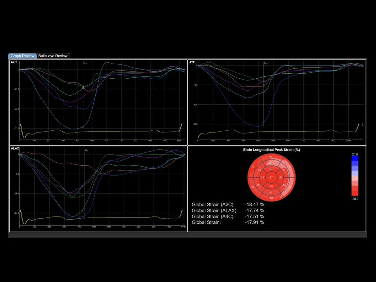
XStrain™ delivers precise global longitudinal strain (GLS) measurements with intuitive visual reports, including bull’s-eye maps and strain curves. This comprehensive assessment aids clinicians in diagnosing cardiomyopathies, chemotherapy-induced cardiotoxicity, and complex structural heart diseases with great confidence, allowing for earlier intervention and improved patient outcomes.
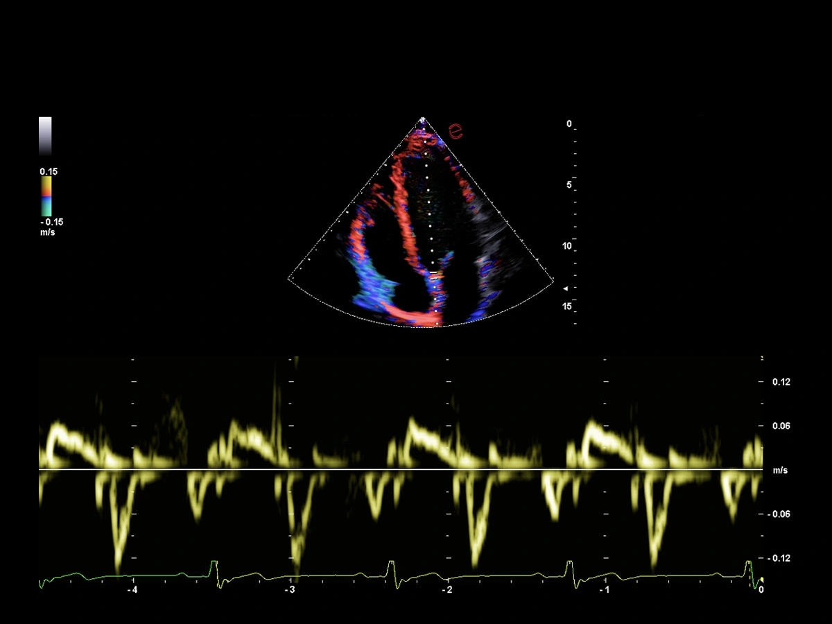
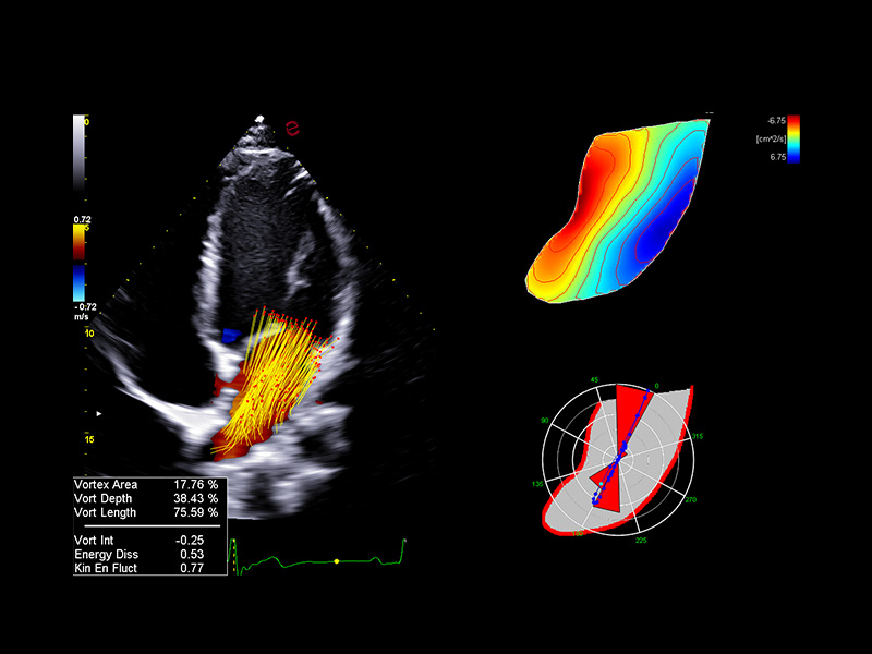
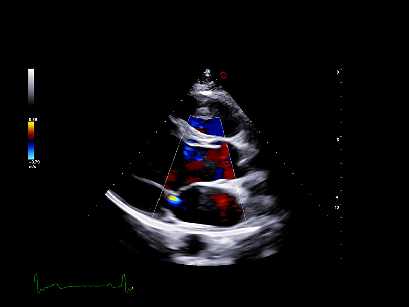
Innovative hemodynamic assessment with HyperDoppler
Esaote’s HyperDoppler technology provides an advanced visualization of intracardiac blood flow dynamics, offering clinicians an intuitive understanding of complex hemodynamics that goes beyond conventional Doppler imaging. By generating real-time vector maps, streamlines, and vortex formations, HyperDoppler enables the detailed assessment of diastolic function, ventricular dyssynchrony, and valvular abnormalities.
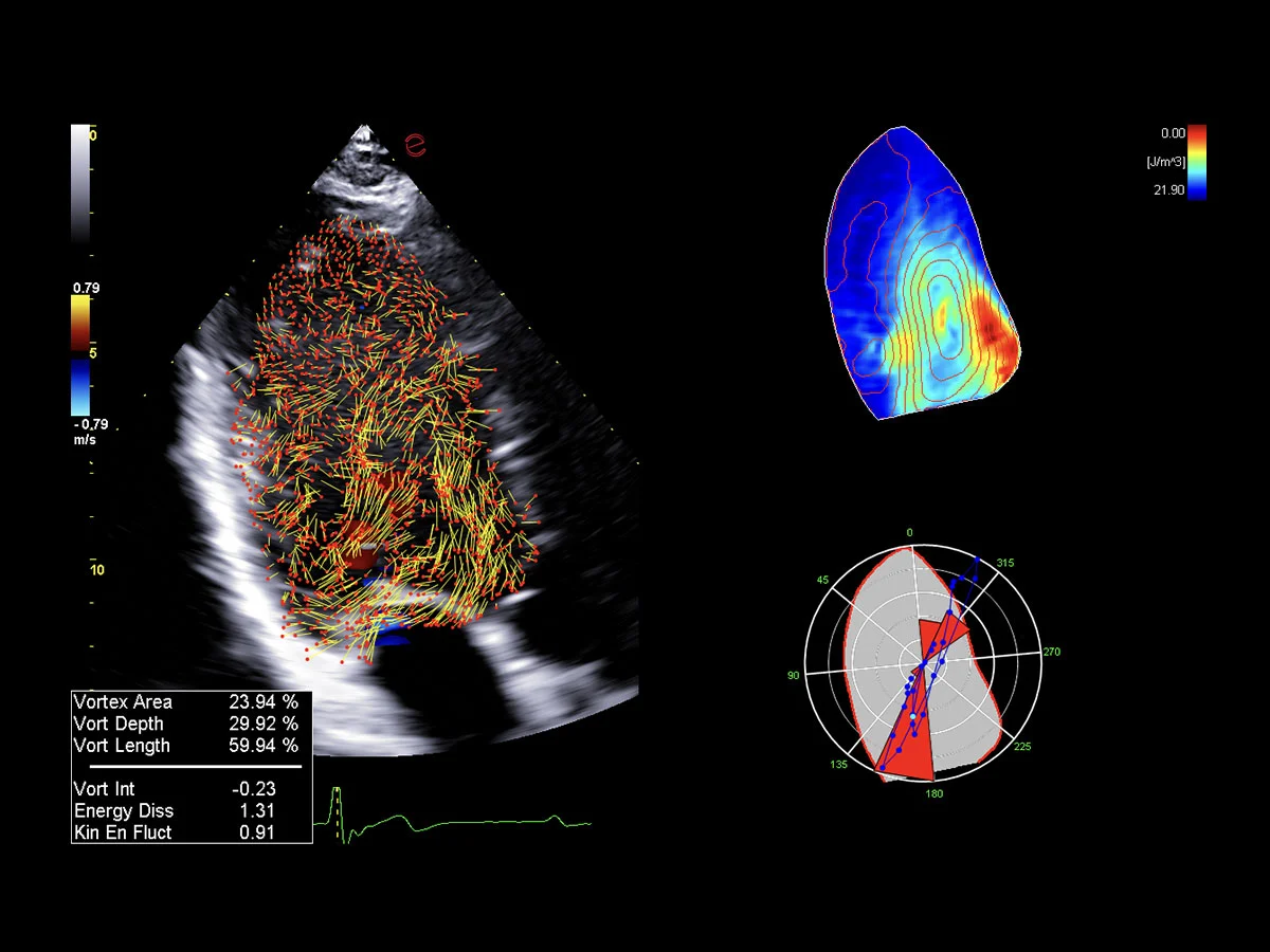
This unique flow visualization supports earlier detection of subtle dysfunction, particularly in challenging cases such as heart failure, prosthetic valve evaluation, and structural heart disease. HyperDoppler’s zero-click, visually rich interface enables faster, more comprehensive exams, allowing cardiologists to make more informed clinical decisions.
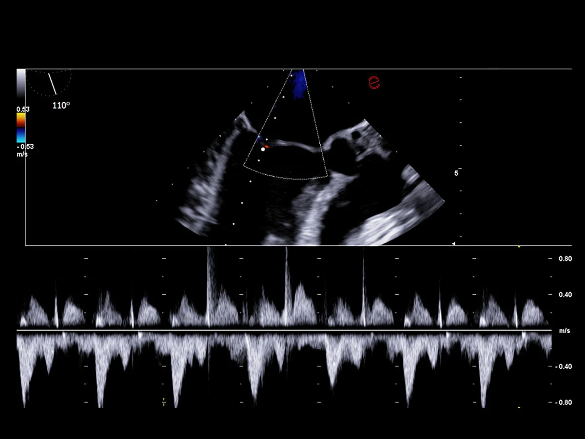
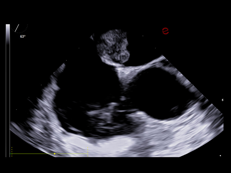
High-performance transesophageal echocardiography probe quality
Esaote’s TEE probe is designed to ensure exceptional image quality and diagnostic versatility, offering detailed visualization of posterior cardiac structures when transthoracic access is limited. It enables precise assessment of the left atrium, appendage, cardiac valves, and thoracic aorta, providing vital information in complex scenarios, such as endocarditis, suspected thrombus, or intraoperative monitoring. Its ergonomic design ensures patient comfort and optimal control during probe manipulation, and its seamless integration with Esaote systems supports real-time guidance for structural interventions and critical diagnostics.
Clinical images
Ultrasound systems for Cardiovascular
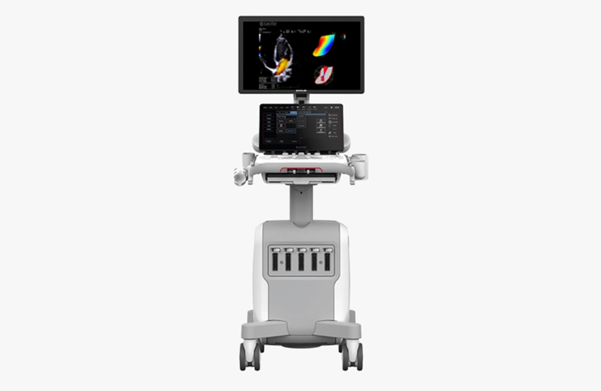
MyLab™E80
Advanced ultrasound platform engineered to provide unparalleled expertise in examinations.
Advanced ultrasound platform engineered to provide unparalleled expertise in examinations.
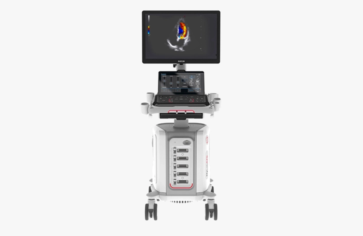
MyLab™A70
Advanced system designed to deliver advanced technologies with the simplest workflow.
Advanced system designed to deliver advanced technologies with the simplest workflow.
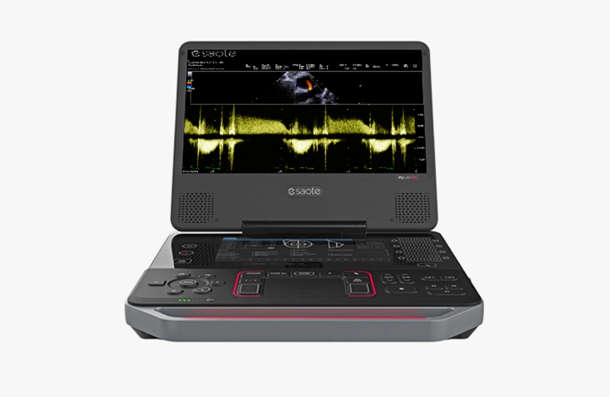
MyLab™C30
Truly compact ultrasound device which combines cutting-edge aesthetics with an efficient user experience.
Truly compact ultrasound device which combines cutting-edge aesthetics with an efficient user experience.
Technology and features are system/configuration dependent. Specifications subject to change without notice. Information might refer to products or modalities not yet approved in all countries. Product images are for illustrative purposes only.
For further details, please contact your Esaote sales representative.
