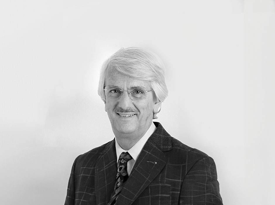
Luigi Solbiati, Busto Arsizio, Varese, Italy, 1952.
Professor of Radiology at Humanitas University (Rozzano – Milan) and consultant interventional radiologist at Humanitas Research Hospital.
Research reveals possible new avenues to the physician, who highlights the boundaries of usefulness to research. Procedures are then revamped and intuitions that may become science are tested. Towards less and less invasive medicine.
As the son of a doctor, who was passionate about photography and cinematography and had chosen radiology (at that time really in its infancy) as his own specialty, I chose to follow the same path for the same reason: a passion for images. I loved the idea of being able to see what the eye could not inside the human body and of trying to correlate those images to each patient’s medical history and clinical data. I, too, chose to become a radiologist (or rather a diagnostic imaging specialist) before I had even completed my medical studies. Within a few years, before university, I went from helping my father and the radiology technicians to develop and fix the x-rays in the darkroom – automatic film processors did not yet exist – to experiencing firsthand the most incredibly rapid and fantastic evolution of diagnostic imaging, probably the medical specialty that has undergone the greatest evolution in the last 50 years, made possible by technology and – even more so – the introduction of computers.
I refer to the birth and stormy development of the three diagnostic methods that revolutionized the art of “looking inside the body”: ultrasound, computed tomography, and magnetic resonance imaging. For all three – thanks to growing technological advancement and the continuous improvement of image quality through constant interaction and comparison between radiologists, engineers, and technicians – there has been a gradual “simplification” in obtaining images, in an increasingly shorter time, and with less need for patient “collaboration”. It is easy to remember how the first manual ultrasound scanners required 10 to 12 minutes and perfect cooperation from the patient in order to obtain an adequate study of the liver, while today a minute or two is more than enough, even with uncooperative patients. Or how the time required for a computed tomography (CT) scan of the abdomen went from several minutes to 3 or 4 seconds with any type of patient, or under 2 seconds when scanning the heart. One of the main complexities solved by technology was the compression of space and time.
While it is true that today’s imaging technology can reveal increasingly precise details, it is also true that, at the end of each diagnostic procedure, a report must be drawn up by a radiologist, who is responsible for the diagnosis.
The medical act will always remain the basis of any action concerning the patient. The machine makes suggestions and shows a universe of options, but it is always a person who validates the next medical action taken on another person. The physician has expertise and sensitivity to context that goes beyond data, even when aided by technology. Accuracy in the collection of the patient’s history (often initiating emotional elements that must be skilfully managed) and comparison with data and results of procedures performed by other specialists are elements that only the physician can manage. This, however, may not allow even the best radiologist to visualize, describe, and interpret elements now revealed by increasingly sophisticated technology. The awareness of these limitations and, in parallel, the incredible development of information technology, has led to the introduction of Artificial Intelligence (AI) into clinical practice, the main benefit of which is how it puts extremely large case libraries at our disposal. The enormous amount of information that can be processed allows cross-simulations and a better highlighting of elements of difference with respect to pictures considered normal, facilitating the identification of pathologies of uncertain visualization even to experienced radiologists, and providing them with an invaluable aid in the formulation of the diagnosis.
AI currently has important clinical applications in mammography, direct radiography, and CT of the chest, as well as in ultrasound of superficial tissues (thyroid and breast in particular), and will soon open up new and interesting applications for us.
Once again, what is the purpose of AI? To simplify. The main limitation to the expansion of these libraries is the complex regulations in various countries that concern the privacy of the patient and make it complicated to circulate diagnostic images. Simplifying these regulations would enable AI to develop significantly.
A further fantastic step that technological progress has made possible is support for the physician not only in the observation of the problem but also in its treatment, guiding needles, catheters, and other mini-instruments inside the body to perform precise interventions “under cover”, without the need for surgical openings. The future of medicine envisions a body that is less and less violated, for both diagnosis and treatment. When, back in 1982 at Busto Arsizio Hospital, we thought of injecting a small amount of dehydrated alcohol into a large parathyroid adenoma in the neck of a patient deemed inoperable in an attempt to achieve sclerosis of the blood vessels in the mass, we certainly did not think that apparently “simple” procedure, made possible by the real-time ultrasound and the precise visual control of the position of the needle inside the body, would open the way to minimally invasive therapies. We simply followed the path embarked on years earlier by our angiologist colleagues, who injected the same type of alcohol into varicose veins to achieve sclerosis without resorting to surgery.
The winning idea was to apply the same treatment inside a tumor, gradually controlling the effect on the target tissue. When, shortly afterwards, Dr. Livraghi at the Vimercate Hospital thought to use the same minimally invasive treatment for hepatocarcinomas in cirrhotic patients – pathologies with high local recurrence in very delicate subjects – he founded the fourth pillar of oncology, interventional oncology, which has established itself alongside medical, surgical, and radiation therapy . Once again, simplicity, coupled with the development of technology, had proven its effectiveness. Today, minimally invasive therapies occupy even greater therapeutic spaces, thanks to increasingly refined and high-performance methods (radiofrequency, microwaves, laser, cryotherapy, chemoembolization, radioembolization, etc.) conducted by increasingly precise guidance technologies.
Esaote has truly played an absolute leading role in this field, as the first company in the world to believe in fusion in the interventional room of a real-time method, ultrasound, with previously captured static and broad exploration methods (CT, magnetic resonance imaging - MRI, CT-PET) in order to precisely reach targets that are partially or not at all visualizable with ultrasound, and then to proceed to their minimally invasive treatment. There are, however, situations of pathology in organs that are not visible by ultrasound (lung, bone, etc.) or in patients not suitable for image fusion. In these, the guidance of minimally invasive therapies can only be performed using CT, resulting in significant radiation exposure, not so much for the patient, but for the operators who must frequently perform these procedures.
These situations are also finding a simple solution in the shape of augmented reality, that is, the precise real-time overlay of physical reality with virtual reality, previously obtained through CT or MRI examinations, and observed through glasses that provide the operator with 3D visualization of organs and targets. In addition, by linking the operator’s glasses with external monitors, which can be in the same examination room or at greater distances, students, residents, and junior staff will be able to observe the investigation as accurately as they would if they were at the operator’s side.
In conclusion, the increasingly sophisticated integration of medical and research experiences from different parts of the globe is exponentially creating opportunities to achieve a broad view and find solutions that will simplify the lives of physicians and patients. Technological research enables us to reveal many things about the human body and how it functions, making us more and more aware that we can increase our potential for action, and revamping procedures so that they are increasingly less invasive. The opportunity is thus created to imagine new trajectories, offering us the space (both in terms of time and psychologically) to evaluate alternatives, including in an emergency, and to test our intuition, even outside the box.

