Liver Ultrasound
Esaote Liver Ultrasound technologies provide a complete solution to assist with the detection, monitoring and treatment of hepatic lesions and clinical liver conditions, with a multiparametric approach to support integrated diagnosis and therapy guidance.
Esaote provides an easy and intuitive liver ultrasound workflow to enhance your performance and confidence, even for the most challenging patient, with automatic tools such as eScan, easyMode and easyColor.
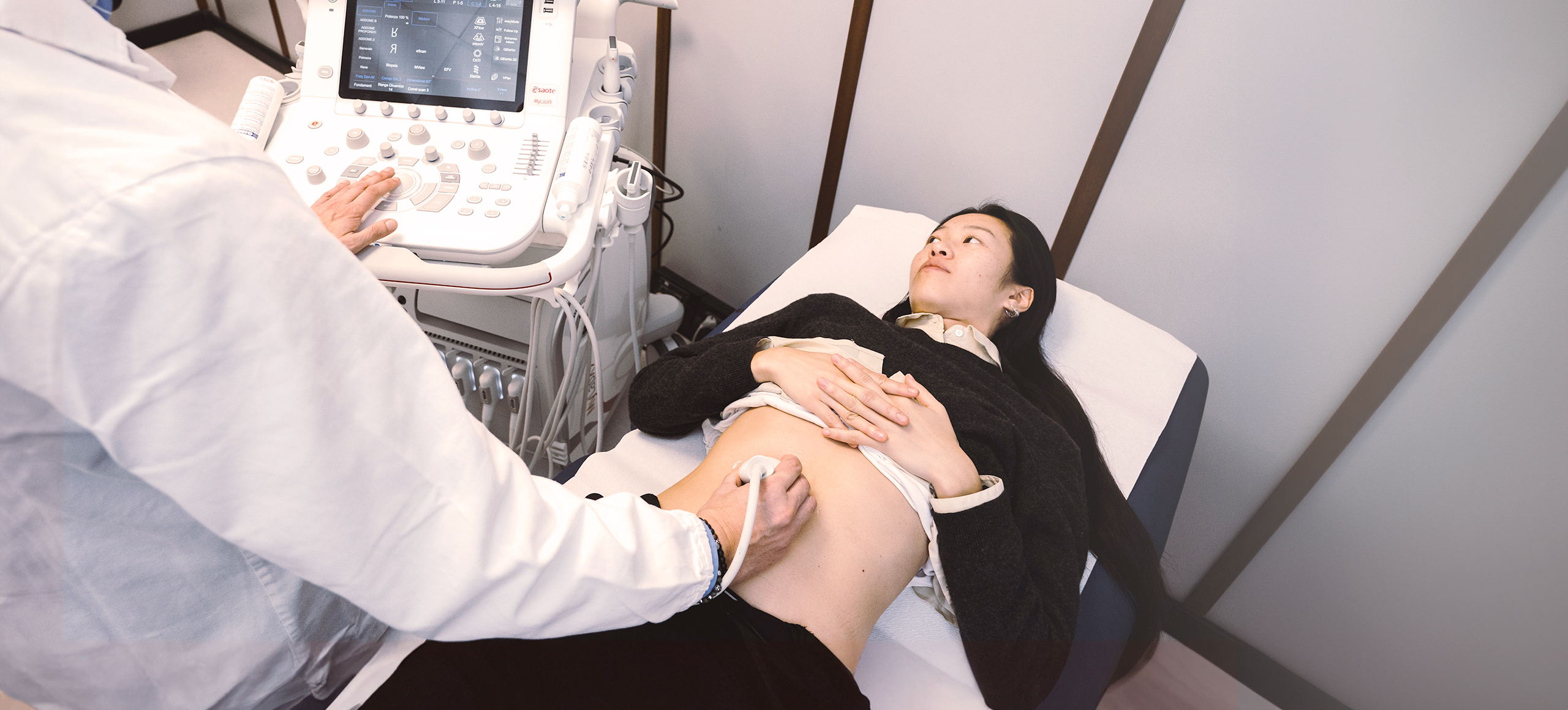
An ultrasound examination may be the first screening conducted to evaluate the condition of the liver, other than blood tests. This technique can be used in association with other imaging modalities, such as CT or MRI, to obtain further information.
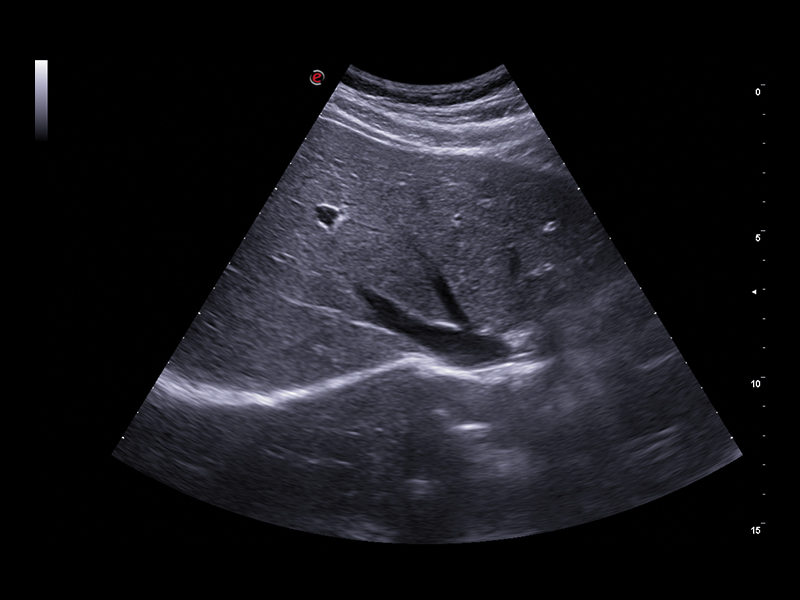
Ultrasound is particularly useful for differentiating solid masses from fluid-filled ones, and can also evaluate diffuse liver diseases, such as fatty liver or hepatitis. Nowadays, extended connectivity and the multimodality approach are opening new horizons in radiology imaging, where ultrasound devices may play a central role in Cross-Modality Imaging.
INTERVIEWS
Testimonials
![]() VIDEO
VIDEO
US approach to patients with liver diseases: practical demonstrations
Prof. Fabio Piscaglia
![]() VIDEO
VIDEO
Liver total approach: clinical case studies (part 1)
Prof. Dr. Dirk-André Clevert
![]() VIDEO
VIDEO
Liver total approach: practical demonstrations (part 2)
Prof. Dr. Dirk-André Clevert
Easy management of challenging patients
Designing its devices by focusing on the customer experience, Esaote has developed smart tools to optimise the image quality in real-time. EasyMode and easyColor automatically adjust the B-mode and color Doppler parameters according to the scanned area and the type of image chosen by the doctor.
Other automatic optimisation tools, such as eScan and eDoppler support you in more rapidly acquiring images with optimal quality, particularly in challenging patients. XCrystal technology developed by Esaote on the convex array enables ultra clarity in very deep and difficult areas while delivering outstanding resolution.
Combining the expertise of Esaote in liver ultrasound through signal management and the zero-click workflow might increase your confidence in the daily screening.
Liver quantification functions
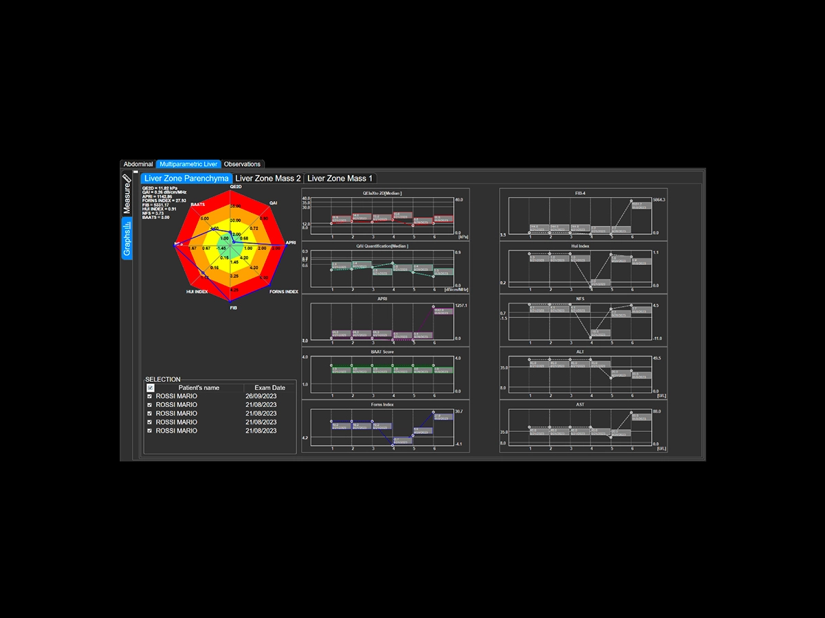
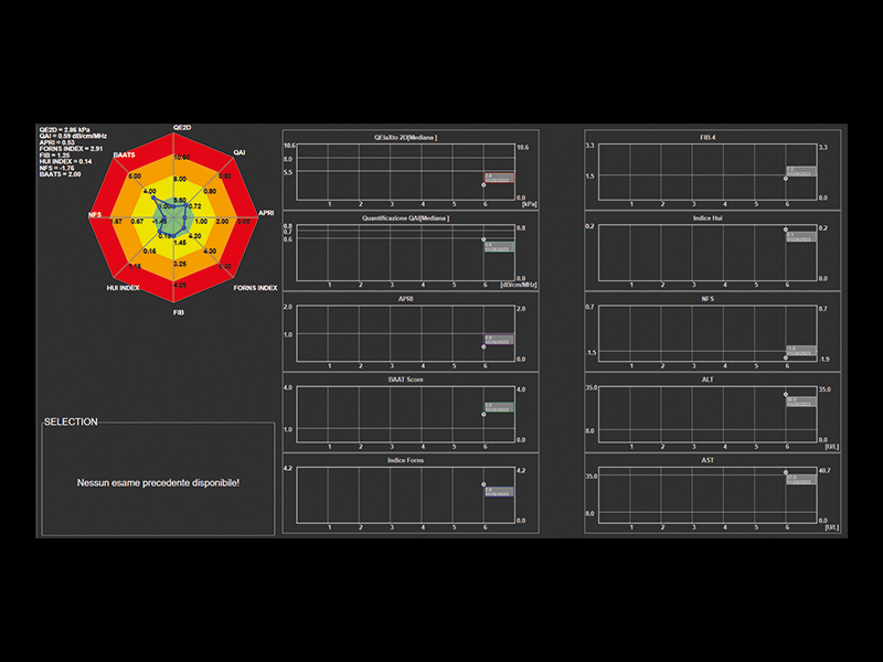
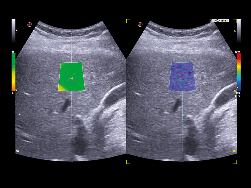
Esaote offers a comprehensive real-time multiparametric package to assess the liver tissue in a non-invasive way. QElaXto 2D, Esaote’s Shear Wave Elastosonography (SWE) technique enables stiffness mapping and provides a quantitative assessment of liver fibrosis.
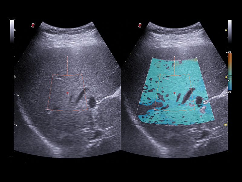
A strong rejection algorithm associated with a dispersion map increases the quality and reliability of the measurements. QAI (Q Attenuation Imaging) is an ultrasound technique for the visualization and quantification of the attenuation along the liver depth provided by Esaote, to support the evaluation of fatty liver tissue and help in the steatosis assessment. The data are computed in a clear multiparametric report with a spider or bar-graph representation to obtain an overview of the liver condition at a glance.
Lesions characterisation
The characterisation of the lesions is essential in the global approach to follow-up and therapy options.
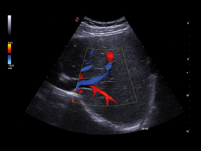
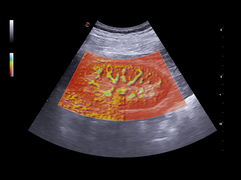
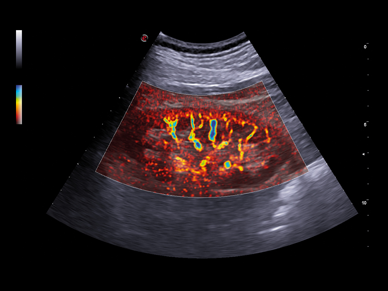
- microV: latest Esaote technology for microvascularisation visualisation with an elevated degree of sensitivity even in very small vessels and with slow flow detection. It enables advanced hemodynamic evaluation of lesions even in deep areas of the liver, thanks to exclusive filters that enhance only blood signals.
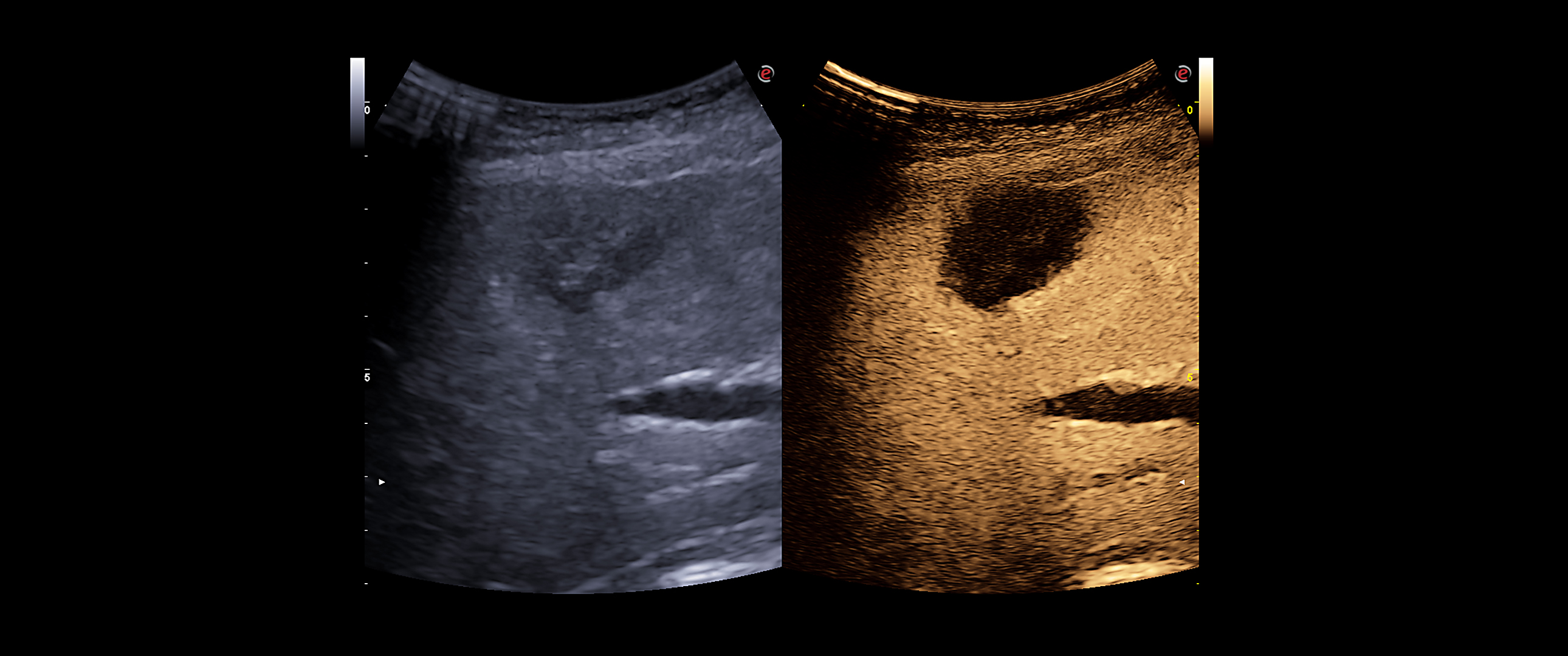
- CnTI™ Clear (Contrast Tuned Imaging): Contrast Enhanced Ultrasound (CEUS) Imaging of Esaote, CnTI™ Clear, detects organ perfusion through contrast agent enhancement to support liver and abdominal lesion characterisations. Based on cutting-edge software, CnTI™ Clear is able to detect low levels of microbubbles, especially in the early arterial phase, while it will ensure their longevity, to give you accurate information on how the lesion is reacting.
- Q-Pack: additionally, Q-Pack is an advanced feature that monitors and quantifies, in graph form and as a function of time, the hemodynamic information detected within a selected ROI, using methods such as CEUS or Doppler Mode.
Liver Interventional procedures
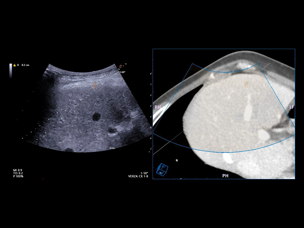
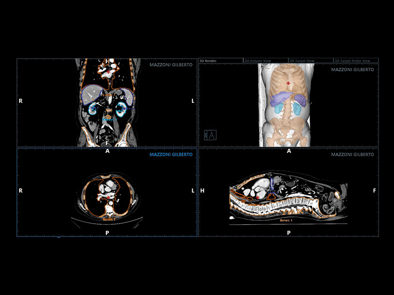
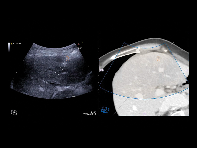
Virtual Navigator (VNav), as the most advanced Esaote Fusion Imaging technology supported by Artificial Intelligence, is opening new horizons in cross-modality liver imaging. Virtual Navigator provides a very simple and active coupling of ultrasound examination with a second Dicom modality imaging (MRI/CT/Pet-CT) dataset as a reference. It enables real-time navigation to benefit from both modalities to increase confidence and accuracy throughout interventional procedures on the liver.
Virtual Navigator will bring you particular support when you face complicated liver pathologies or conditions during your procedures, particularly in the following cases:
- Lesions which are better identified with CT, MRI, and PET or slightly visible in US
- Lesions which are only visible during arterial phase enhancement
- Follow-up of local tumours after ablation or resection
- New lesions in subsequent follow-up surgery or ablation.
- Hidden lesions during treatments (gassed out US).
- Composite ablations requiring multiple needle insertions.
- Complex geometries or difficult treatment planes to identify a safe pathway to the target, such as a difficult US “window”, or complex angle of insertion.
Virtual Navigator, supported by A.I., simplifies abdominal fusion thanks to the exclusive AutoSync function that enables automatic registration based on 3D camera technology. As a historical expert in fusion imaging, Esaote offers you a comprehensive package of tools to significantly reduce the complexity of interventional procedures and increase confidence in the information obtained with your ultrasound device.
- Augmented reality visualisation of the abdominal organs
- Automatic movement correction
- Needle tracking
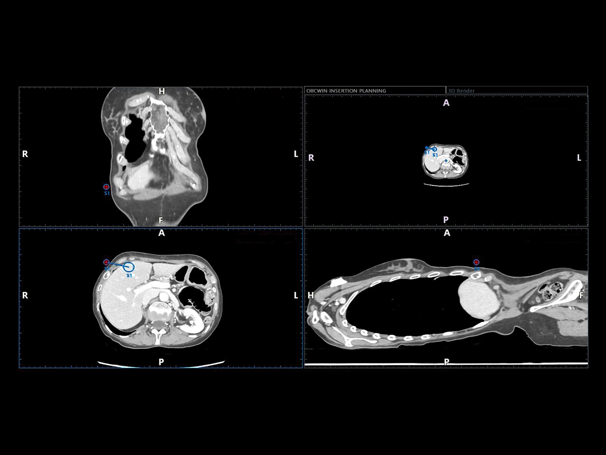
- Advanced planning software
- CEUS, elastosonography, microV compatibility
- Automatic registration via omniTRAX™
Product images are for illustrative purposes only. For further details, please contact your Esaote sales representative.
omniTRAX is a trademark of CIVCO Medical Solutions.
MyLab is a trademark of Esaote spa. CnTI™: The use of Contrast Agents in the USA is limited by FDA to the left ventricle opacification and to characterization of focal liver lesions.
Clinical Images

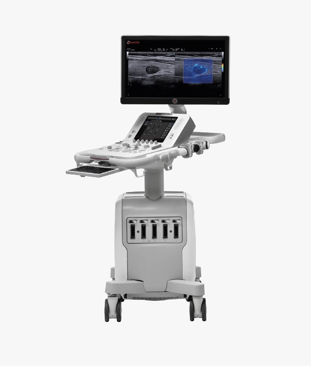
Ultrasound systems for Liver Care
Technology and features are system/configuration dependent. Specifications subject to change without notice. Information might refer to products or modalities not yet approved in all countries. Product images are for illustrative purposes only.
For further details, please contact your Esaote sales representative.
