Whole body imaging
As a pioneer in healthcare technology and MRI solutions, we are dedicated to providing advanced imaging tools that empower clinicians in delivering exceptional patient care. Our specialized MRI solutions offer comprehensive diagnostic capabilities for a wide range of conditions, from abdominal disorders to neurological pathologies. With cutting-edge imaging technology, we enable the detailed visualization of healthy and pathological tissues, supporting accurate diagnoses and informed decision-making.
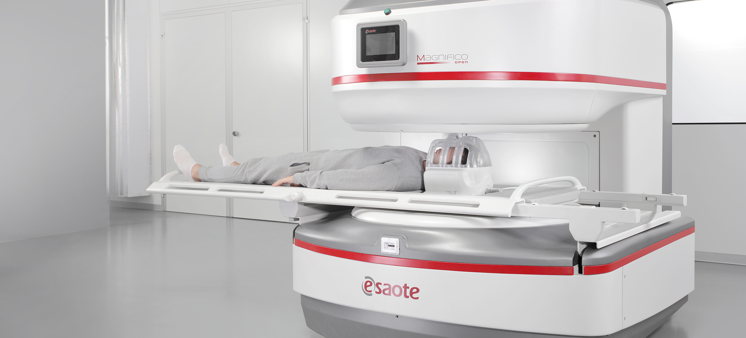
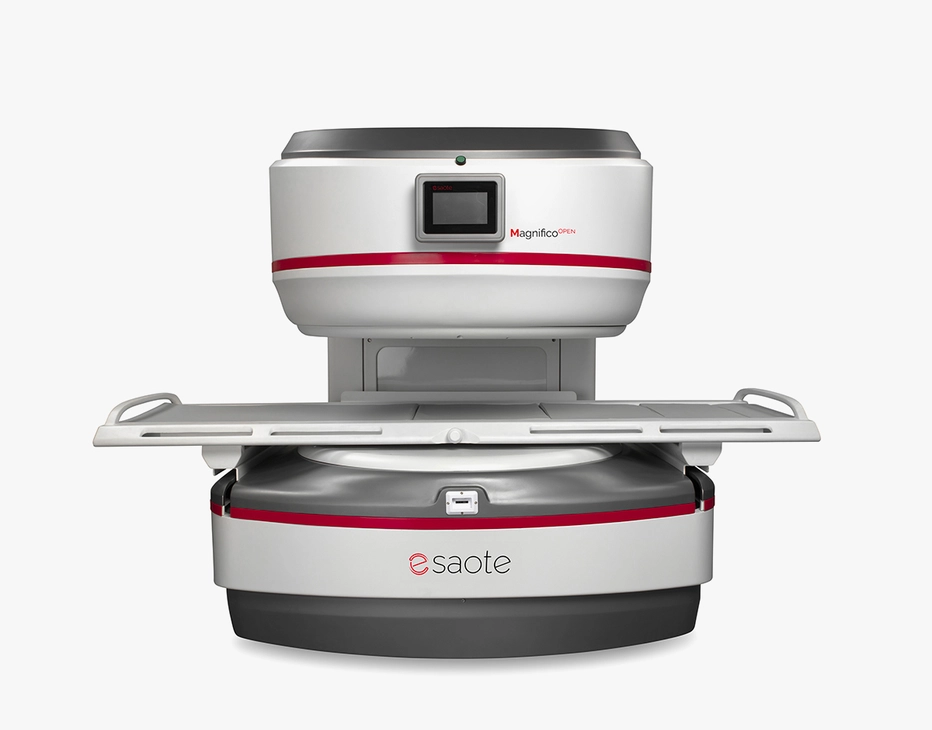
Whether for abdominal imaging or neuroimaging, our systems are designed to combine clinical efficiency with patient comfort. From initial screenings to advanced evaluations, features such as diffusion imaging and angiographic sequences enhance diagnostic potential, even in compact and accessible settings. Focused on innovation, precision, and reliability, we are committed to supporting healthcare professionals in achieving excellence through state-of-the-art MRI technologies.
- Patient-friendly design
Open configuration that minimizes claustrophobia, particularly beneficial for patients prone to anxiety.
- Safety and accessibility focus
Low-field MRI enhances safety but also widens accessibility, making the MRI setting more comfortable and open to all.
Neuroimaging MRI
The Time-Of-Flight (TOF) effect in MRI arises due to the blood flow between the RF pulses. The TOF effects in gradient-echo imaging appear as a signal hyperintensity due to the in-flow of fresh blood that has not experienced any prior RF pulses. Nonetheless, Esaote implemented innovative techniques to increase the image resolution and decrease sensitivity to the effects of flow saturation. The User Interface also includes Maximum Intensity Projection (MIP) software to proficiently reconstruct MR Angiograms detail vascular area of interest.
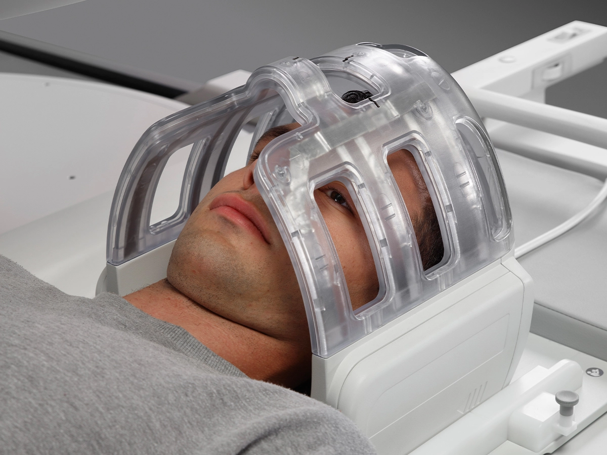
Low-field MRI brain examination
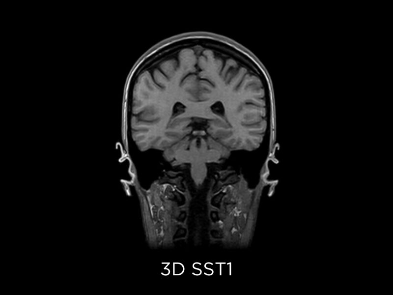
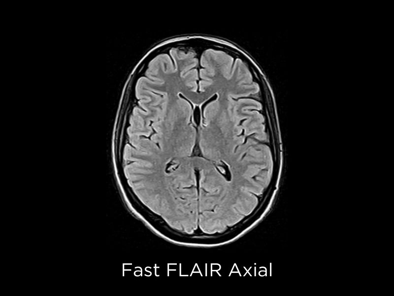
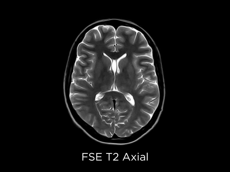
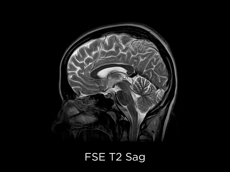
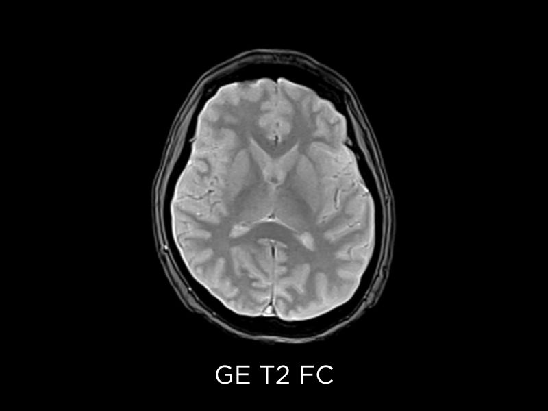
Generally speaking, low-field systems are used for screening purposes and the first investigation of common cerebral diseases, such as trauma, headache, and cognitive impairment. Even at low field, Diffusion imaging and Angiographic sequences provide an extra value enriching the diagnostic capabilities in the same compact and tolerable setting.
Neuroimaging clinical images
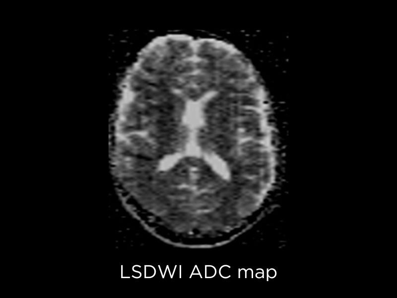
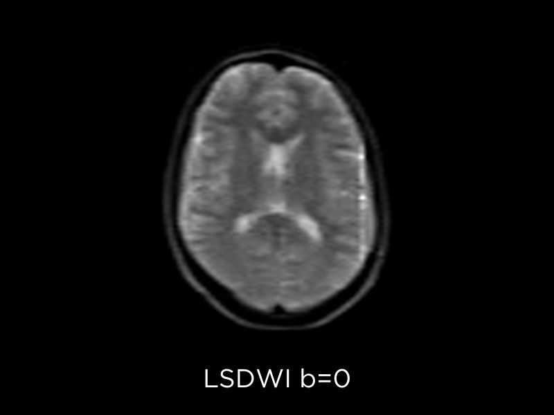
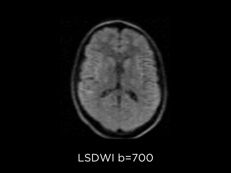
Diffusion Weighted Imaging (DWI) is a fundamental sequence to detect brain tumors, their classification and grading and monitoring. On Magnifico Open, Esaote implemented the DWI sequence with line scan technology1: this technique is based on spin echo sequences and has reduced susceptibility to static field inhomogeneities than echo planar imaging (EPI). In addition, it does not require enhanced gradient hardware and offers easy installation and reduced power consumption.
DWI provides important clinical value especially in the assessment of Cerebral Infarction and Stroke, Neoplasia, Emphysema, Toxic and Demyelinating pathologies.
Neuroimaging clinical images
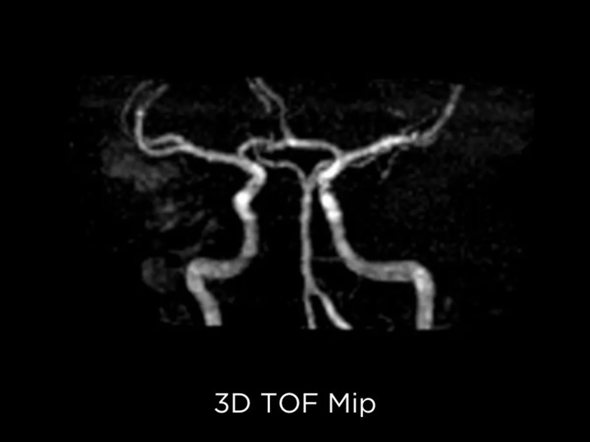
TOF sequences
Time of Flight angiography (TOF) is aimed at visualizing flow within vessels, even without contrast injection (MRA). 2D TOF is more sensitive to slow flow and it is more useful to assess long vessels whilst 3D TOF usually is preferred in the study of tortuous vessels (I.e. Circle of Willis)3. These sequences are crucial in the assessment of several conditions such as intracranial aneurysm and vascular stenosis.
The Time-Of-Flight (TOF) effect in MRI arises due to the blood flow between the RF pulses. The TOF effects in gradient-echo imaging appear as a signal hyperintensity due to the in-flow of fresh blood that has not experienced any prior RF pulses. Nonetheless, Esaote implemented innovative techniques to increase the image resolution and decrease sensitivity to the effects of flow saturation. The User Interface also includes Maximum Intensity Projection (MIP) software to proficiently reconstruct MR Angiograms detail vascular area of interest.
Abdominal Examination*
With a focus on innovation, reliability, and precision, we continue to support medical professionals in achieving excellence in patient outcomes through advanced imaging technologies.
* Not available for sale in USA
This feature might be subject to regulatory requirements, therefore prohibiting its sale in certain countries. For further details, please contact your Esaote sales representative.
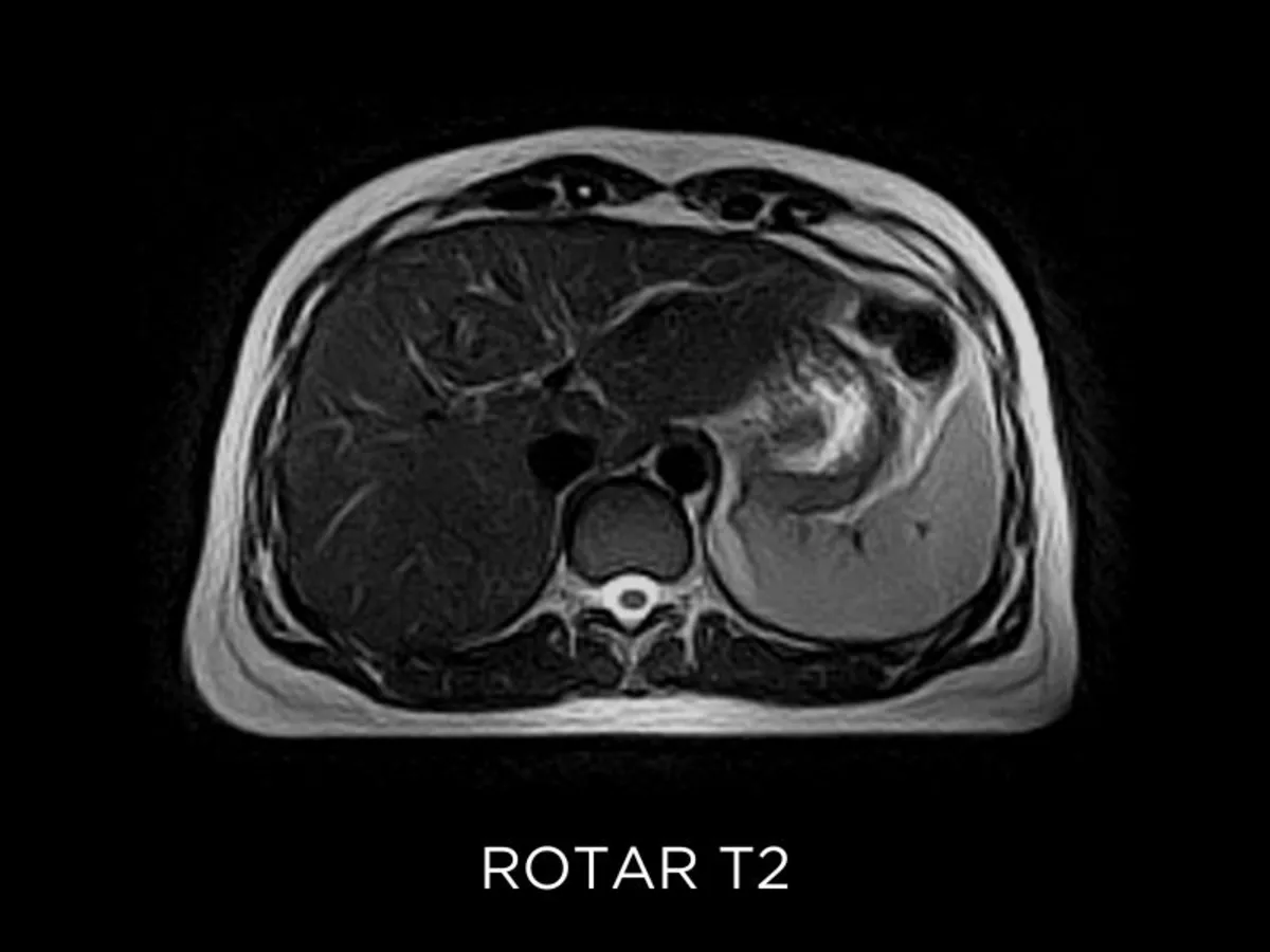
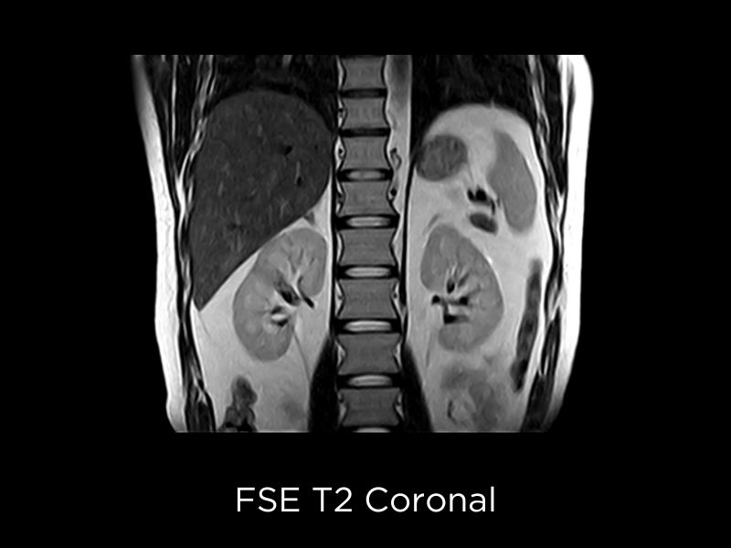
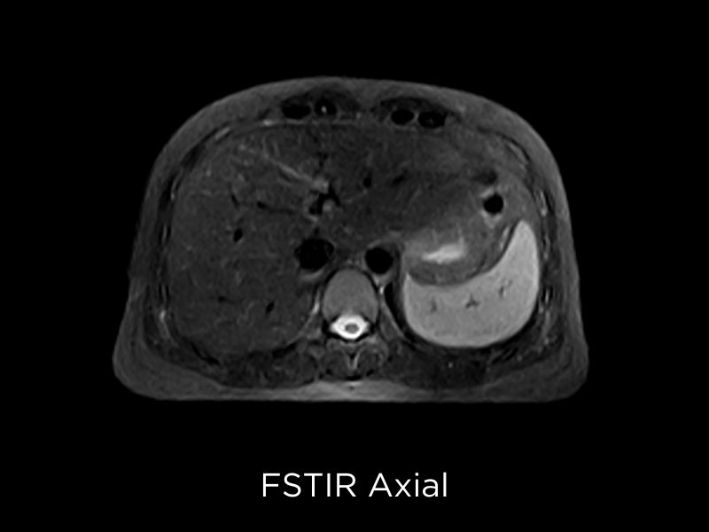
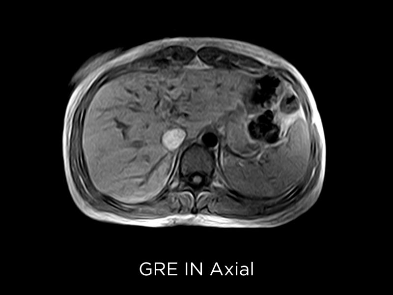
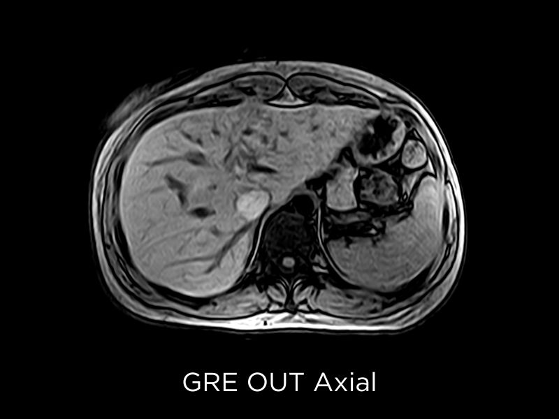
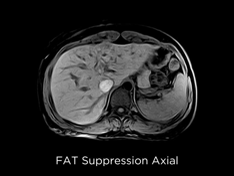
Upper abdomen investigations can be conducted using three types of coils:
- Small Spine Coil,
- Large Spine Coil
- and Body Coil.
The respiratory belt, featuring an elastic texture and dedicated clasp, can be adjusted to fit different patient sizes. The user interface allows for respiratory gating control and supports breath-hold management with options for inspiratory or respiratory triggering.
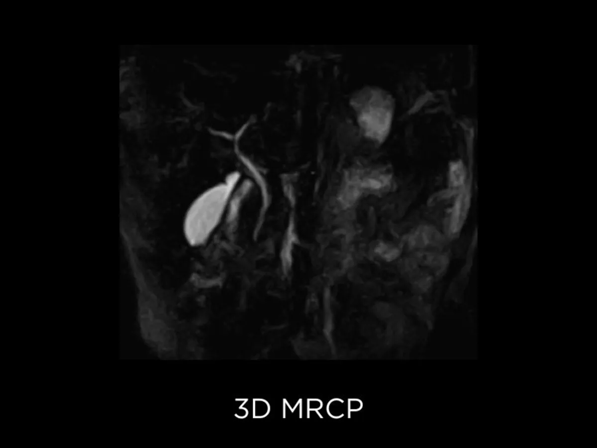
Available Techniques
Available techniques include:
- ROTAR: in FSE sequences, it reduces moving artifacts on free-breathing acquisitions.
- ROPE: enhances triggering management in non-FSE sequences by minimizing signal dependency on the patient’s breath.
- FSE 3D: provides high SNR and contrast for thin-slice MRCP imaging.
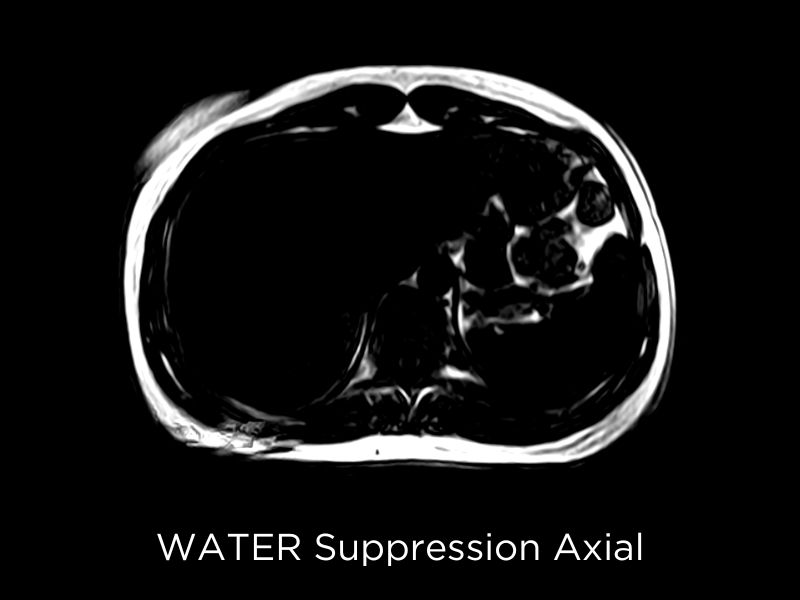
Abdominal imaging clinical images
Sources
- MRI Exam-Specific Parameters: Head and Neck Module (Revised 4-6-2022)
https://accreditationsupport.acr.org/support/solutions/articles/11000061019-mri-exam-specific-parameters-head-and-neck-module-revised-4-6-2022- - Drake-Pérez, M., Boto, J., Fitsiori, A. et al. Clinical applications of diffusion weighted imaging in neuroradiology. Insights Imaging; 9, 535–547 (2018). https://doi.org/10.1007/s13244-018-0624-3
- Gudbjartsson, H., Maier, S.E., Mulkern, R.V., Mórocz, I.Á., Patz, S. and Jolesz, F.A. (1996), Line scan diffusion imaging. Magn. Reson. Med., 36: 509 519. https://doi.org/10.1002/mrm.1910360403
- D. Saloner; The AAPM/RSNA physics tutorial for residents. An introduction to MR angiography; RadioGraphics 1995 15:2, 453-465
- Castillo, M., Camilo Márquez, J. and José Medina, F. (2014). Time-of-Flight Magnetic Resonance Angiography (TOF MRA) and MRV: Clinical Applications. In Vascular Imaging of the Central Nervous System (eds J.N. Ramalho and M. Castillo). doi.org/10.1002/9781118434550.ch9
Related system for Whole body imaging*
Technology and features are system/configuration dependent. Specifications subject to change without notice. Information might refer to products or modalities not yet approved in all countries. Product images are for illustrative purposes only.
For further details, please contact your Esaote sales representative.
* Not available for sale in USA
This feature might be subject to regulatory requirements, therefore prohibiting its sale in certain countries. For further details, please contact your Esaote sales representative.
